|
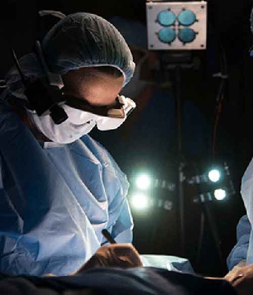
High-tech glasses developed at Washington University School of Medicine in St. Louis may help surgeons visualize cancer cells, which glow blue when viewed through the eyewear. The wearable technology, so new it's yet unnamed, was used during surgery for the first time today at Alvin J. Siteman Cancer Center at Barnes-Jewish Hospital and Washington University School of Medicine. Cancer cells are notoriously difficult to see, even under high-powered magnification. The glasses are designed to make it easier for surgeons to distinguish cancer cells from healthy cells, helping to ensure that no stray tumor cells are left behind during surgery. "We're in the early stages of this technology, and more development and testing will be done, but we're certainly encouraged by the potential benefits to patients," said breast surgeon Julie Margenthaler, MD, an associate professor of surgery at Washington University, who performed today's operation. "Imagine what it would mean if these glasses eliminated the need for follow-up surgery and the associated pain, inconvenience and anxiety." Current standard of care requires surgeons to remove the tumor and some neighboring tissue that may or may not include cancer cells. The samples are sent to a pathology lab and viewed under a microscope. If cancer cells are found in neighboring tissue, a second surgery often is recommended to remove additional tissue that also is checked for the presence of cancer. The glasses could reduce the need for additional surgical procedures and subsequent stress on patients, as well as time and expense. Margenthaler said about 20 to 25 percent of breast cancer patients who have lumps removed require a second surgery because current technology doesn't adequately show the extent of the disease during the first operation. "Our hope is that this new technology will reduce or ideally eliminate the need for a second surgery," she said. The technology, developed by a team led by Samuel Achilefu, PhD, professor of radiology and biomedical engineering at Washington University, incorporates custom video technology, a head-mounted display and a targeted molecular agent that attaches to cancer cells, making them glow when viewed with the glasses. In a study published in the Journal of Biomedical Optics, researchers noted that tumors as small as 1 mm in diameter (the thickness of about 10 sheets of paper) could be detected. Ryan Fields, MD, a Washington University assistant professor of surgery and Siteman surgeon, plans to wear the glasses later this month when he operates to remove a melanoma from a patient. He said he welcomes the new technology, which theoretically could be used to visualize any type of cancer. "A limitation of surgery is that it's not always clear to the naked eye the distinction between normal tissue and cancerous tissue," Fields said. "With the glasses developed by Dr. Achilefu, we can better identify the tissue that must be removed." In pilot studies conducted on lab mice, the researchers utilized indocyanine green, a commonly used contrast agent approved by the Food and Drug Administration. When the agent is injected into the tumor, the cancerous cells glow when viewed with the glasses and a special light. Achilefu, who also is co-leader of the Oncologic Imaging Program at Siteman Cancer Center and professor of biochemistry and molecular biophysics, is seeking FDA approval for a different molecular agent he's helping to develop for use with the glasses. This agent specifically targets and stays longer in cancer cells. "This technology has great potential for patients and health-care professionals," Achilefu said. "Our goal is to make sure no cancer is left behind." Dr. Achilefu has worked with Washington University's Office of Technology Management and has a patent pending for the technology. The research is funded by the National Cancer Institute (R01CA171651) at the National Institutes of Health (NIH).
|
透過一副特制的高科技眼鏡,醫(yī)生小心切除閃著藍(lán)光的癌變組織……當(dāng)?shù)貢r(shí)間2月10日,一臺(tái)特殊的外科手術(shù)在美國密蘇里州圣路易斯市一家醫(yī)院里進(jìn)行,借助這項(xiàng)剛剛問世的可視技術(shù),原本幾不可見的癌細(xì)胞變得無所遁形。 據(jù)美國媒體報(bào)道,這副能夠“看見”癌細(xì)胞的眼鏡由華盛頓大學(xué)(W U)放射學(xué)和生物醫(yī)學(xué)工程學(xué)教授塞繆爾?阿基里弗教授領(lǐng)隊(duì)研發(fā)成功,在理想狀態(tài)下,它可以幫助外科醫(yī)生在手術(shù)時(shí)一次性徹底切除所有癌變組織。 眾所周知,即使置于高性能的顯微鏡下,癌細(xì)胞也難以被發(fā)現(xiàn)。但有了這副眼鏡之后,醫(yī)生能夠輕松區(qū)分健康細(xì)胞和癌細(xì)胞,從而確保首次手術(shù)時(shí)不會(huì)遺漏任何癌變組織,進(jìn)行二次切除手術(shù)以“查缺補(bǔ)漏”的可能性也由此大大降低。 據(jù)介紹,使用時(shí),需要先把一種特定的分子藥劑涂抹在腫瘤及其周邊組織上,這種藥劑會(huì)附著于癌細(xì)胞、令其發(fā)出肉眼不可見的光芒,然后,主刀醫(yī)生戴上一個(gè)形似眼鏡的頭盔式顯示器,通過自定義視頻技術(shù),即可看清癌細(xì)胞分布于何處。 “目前,我們尚處于研究初期,未來會(huì)進(jìn)行更多的改進(jìn)和測試工作。”乳腺外科醫(yī)生朱莉?馬格塔勒是10日進(jìn)行手術(shù)的主刀大夫,同時(shí)也是第一個(gè)在實(shí)際操作中使用癌細(xì)胞可視眼鏡的人,“一想到這項(xiàng)新技術(shù)將令病患獲益良多,我們就干勁十足。” 依據(jù)現(xiàn)在的醫(yī)護(hù)流程,外科醫(yī)生手術(shù)時(shí)需要切除腫瘤及其附近組織,而這些組織中可能存在、也可能不存在癌細(xì)胞。隨后,切除下的組織標(biāo)本被送往病理實(shí)驗(yàn)室接受檢驗(yàn),如果在其中發(fā)現(xiàn)癌細(xì)胞,則需進(jìn)行第二次甚至多次手術(shù),直至癌變組織被完全切除。 據(jù)馬格塔勒介紹,依靠現(xiàn)有的技術(shù),無法準(zhǔn)備判定癌變組織的全部范圍,所以大約20%至25%的乳腺癌患者接受首次手術(shù)切除腫瘤后,還需經(jīng)受第二次手術(shù)。 “借助這種癌細(xì)胞可視眼鏡,可以在首次手術(shù)時(shí)一次性切除所有癌變組織。這意味著,沒有必要再進(jìn)行后續(xù)手術(shù),病人也無需承擔(dān)隨之而來的病痛和手術(shù)費(fèi)用。”馬格塔勒希望,這項(xiàng)新技術(shù)能夠降低、甚至完全消除二次手術(shù)。 相關(guān)閱讀 受奧朗德與瓦萊麗分手影響 白宮報(bào)廢300份國宴邀請函 科學(xué)家發(fā)現(xiàn)非洲以外人類最古老的腳印遺跡 男子劫機(jī)欲攪亂冬奧會(huì) 計(jì)劃泡湯反被逮捕 美國警告稱恐怖分子或?qū)λ髌醵瑠W會(huì)發(fā)動(dòng)“牙膏炸彈”襲擊 (信蓮 編輯:玉潔)
|
|
|
